
The Cervical Spine

When you hear someone mention the cervical spine or the cervicals, do you know what they are referring to? If not that’s okay, because we are here to share the knowledge! The cervicals are the bones located at the top of your spine, otherwise known as your neck. It consists of seven bones known as “vertebrae”. The first two vertebrae of your cervical spine are unique in function and shape. The first vertebra (C1), also called the atlas, is a ring-shaped bone that begins at the base of your skull and holds our head upright. Your C2 or second vertebra, also known as the axis, allows the atlas to pivot against it for the side-to-side “no” rotation of your head. C3-C7 are all very similar and just increase in size little by little as we go down the spine. All seven vertebra are connected by a joint called a “facet” that is located at the back of each bone and allows our necks to move forward, backwards, and twisting side to side. The cervical spine is supposed to have a “lordotic” curve meaning that it slightly curves to the front of the body. Your cervical spine is also surrounded by muscles, tendons, and ligaments. Intervertebral discs are positioned between each vertebra to act as “shock-absorbers” from gravity and impact. Your spinal cord also runs through the center of your entire spine, as it sends and receives messages from your brain, which controls all aspects of your body’s functions.
Your cervical spine serves many functions, but the three major ones are:
- Protects your spinal cord- Your spinal cord runs throughout your entire spine through holes called “vertebral foramen” that are the center of each vertebra.
- Supports our heads and allows all movement– The average head weighs 10-13 pounds and is supported by your cervical spine and it also allows for movements such as tilting forward (flexion), tilting backwards (extension), bending from side to side (lateral bend or ear to shoulder), and turning side to side (rotation).
- Providing safe passageways for our vertebral arteries– Our brain needs blood to function, and our vertebral arteries do just that, C1-C6 are the only vertebrae in the entire spine that house tiny holes that create safe passageways for these arteries.
The major muscles that attach to your cervical spine include:
- Sternocleidomastoid (SCM)– There is one on each side of your neck. It runs from behind your ear to the front of your neck where it attaches to your breastbone (sternum) and collarbone. It allows for rotation side-to-side and the tilting of your chin upward.
- Trapezius– A pair of triangular muscles extending from the base of your skull down your cervical and thoracic spine and all the way out to your shoulder blade. They help tilt your head upward/move your neck back, rotating your head right or left, and lifting your shoulder blades.
- Levator scapulae– Attaches to your first four cervical vertebrae and the top of your shoulder blade (scapula). Aids in lifting your shoulder blade, bending your head to the side, and rotating your head.
- Erector spinae– Several muscles make up this group. In the cervical spine, they help with posture, backward neck extension and neck rotation.
- Deep cervical flexors- Run down the front of your cervical spine allowing flexion of your neck forward and they help keep your cervical spine stable.
- Suboccipital muscles– These four pairs of muscles, connect the base of your skull with the top of your cervical spine which, allows for extension and rotation.
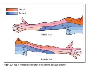
Eight pairs of spinal nerves exit your cervical spine. They’re labeled C1 through C8. They stimulate muscle movement in your neck, shoulders, arms, and hands, and also provide sensation known as dermatome patterns.
- Cervical nerves C1, C2 and C3 control your forward, side to side, and backwards head and neck movements. The C2 nerve will provide sensation to the upper area of your head while C3 gives sensation to the side of your face and the back of your head.
- Cervical nerve 4 controls your upward shoulder motion and is one of the nerves that controls your diaphragm (muscles that helps you breathe and are at the bottom of your rib cage). C4 provides sensation for parts of your neck, shoulders, and upper arms.
- Cervical nerve 5 controls the deltoid musculature of your shoulders and biceps. C5 provides sensation to the upper part of your arm and down to your elbow.
- Cervical nerve 6 controls the extensor wrist muscles and is also involved in the control of your biceps. C6 provides sensation to the thumb side of your forearm and hand.
- Cervical nerve 7 controls your triceps and wrist extensor muscles. C7 provides sensation to the back of your arm into your middle finger.
- Cervical nerve 8 controls your hands and gives sensation to the pinky side of your hand and forearm.
There are many diseases and disorders that can occur within the cervical spine. We will go over some of the more common ones but also touch on some of the less frequent as well.
- Cervical radiculopathy: Occurs when a cervical nerve is pinched by cervical vertebrae. You may experience tingling, numbness, weakness, and pain. Symptoms may remain local or can spread to your entire arm, hand and/or fingers. It is also known as a pinched nerve or cervical nerve compression.
- Degenerative Disc Disease (DDD): Occurs when your intervertebral discs (IVDs) wear down and become very thin.
- Disc herniation: occur when the discs tear or leak from either wear and tear or trauma.
- Osteophytes (bone spurs): Occurs when DDD is present, and the vertebrae start making contact and causes the bone to spur or grow outwards and can impinge on the cervical nerves.
- Cervical spondylosis: Also known as arthritis of the neck. It is age related slow degeneration of your discs and joints.
- Stenosis: Occurs when your spinal canal in the cervical spine area narrows. Less space within your cervical spine reduces the amount of space available for your spinal cord and nerves that branch off the spinal cord. This tightened space can cause your spinal cord or nerves to become irritated, compressed, or pinched.
- Cord compression (myelopathy): Applies when there is pressure on your spinal cord within the cervical area. One of the most common causes is wear and tear on the bones of your spine, a condition called osteoarthritis.
- Trauma injuries (cord injury or spinal fracture): Sustained injuries by a strong force to the neck resulting in an injury directly to the cord or for the cervical vertebrae to fracture which can also in turn injure the cord as well.
- Cervical strain or sprain: These are common and can happen for a multitude of reasons, but these are injuries to your cervical musculature and ligaments/tendons.
There are many ways to assess your possible cervical disease or disorder. A thorough evaluation of ROM, orthopedic testing and palpation can tell us a lot but sometimes we need further evidence and that may require radiographs (X-rays), CT, MRI or even an EMG. X-rays will be best to see bones and the joint spaces while an MRI is best to see soft tissue and disc injuries. A CT is best to see the size and injury to your spinal canal and an EMG is best to test any nerve damage. It is important to note that chiropractic care can help with all of these problems in some way but sometimes we need to refer out for help and that can range from physical therapy to massage and all the way to surgery. If you or anyone you know may be experiencing one of the above issues, please contact us for a further evaluation or if you have any further questions, we would be happy to help ☺